Saturday 25 July 2015

Stem cell transplantation benefits in leukemia

Stem cell transplantation to treat patients with a serious but very rare form of chronic blood cancer called juvenile myelomonocytic leukemia (JMML) improves the condition, finds a study.

Allogeneic hematopoietic stem cell transplantation (HSCT) involves the transplantation of stem cells from a donor, which may be derived from bone marrow, peripheral blood or umbilical cord blood.

The recipient's immune system is usually destroyed with radiation or chemotherapy before the transplantation.

"The lack of transplant-related mortality in the group of children we studied at the Children's Center for Cancer and Blood Diseases suggests that BUMEL (Intravenous Busulfan and Melphalan) may represent a successful (HSCT) high-dose chemotherapy regimen," said lead author Hisham Abdel-Azim from the Children's Hospital Los Angeles.

"It is also possible that administering conventional dose chemotherapy, before HSCT, to patients with more progressive disease may have contributed to the improved outcomes," Abdel-Azim said in the journal Blood.

The study looked at children with JMML who underwent HSCT at Children's Hospital Los Angeles.

All of the patients were alive and in clinical remission.

It is the only reported cure for JMML; however best outcomes of the therapy have shown that only half the patients can be cured from this disease.

There is currently no standard conditioning regimen for children with JMML undergoing HSCT.

Friday 24 July 2015

The Revolution Of Stem Cell Therapy And Facts To Know About It

Stem cells are presently being used for creating functional as well as living tissues for repairing or renewing organs and tissues of human body, which are damaged because of chronic illness, accidents, or aging. Stem Cell therapy has revolutionized the treatment of blood related ailments, especially treatment of children suffering from leukemia. This therapy is also used for the purpose of tissue grafts to cure bone injury, or injury to eye surface or skin. Ongoing research on Stem Cell therapy helps to explore new possibilities to use stem cells for treating many other health conditions.

Taking organ transplants to another level

Stem Cell therapy helps to promote the repairing of damaged or diseased tissues with the use of stem cells. This therapy takes organ transplantation to the next level. In place of organ from a donor, this therapy makes use of stem cells. In times, where huge number of patients has to wait for organ transplantation due to lack of donor, stem cells can lend a hand in their treatment. These cells are grown in labs and are of different types like blood cells, never cells, or cardiac muscle cells. The cells once implanted in the patient can help to repair the damaged muscle or tissue of the body.

Advent of the therapy in India

Treatment and research on Stem Cell therapy is going on in many countries including China, Mexico, Middle East, Thailand, Ukraine, and even in India. Of late, Stem Cell therapy in India has gained popularity and trust. There have many successful cases where Stem Cell therapy has successfully treated spinal injuries, nerve damages, and blood disorders. The country is ready to explore the immense potential of this new treatment method. Earlier, the country has witnessed several stem cell transplants, which is also called bone marrow transplant. Recently, adult stem cells are being tested for treating other conditions.

Limitations to consider

Despite of all the constant research going on in the field of Stem Cell therapy in India and its success stories, there are a few limitations, which one should keep in mind. Only some of the treatments are considered effective and safe presently. The treatment of an ailment depends on the type of stem cell being used, as different kind of stem cells are used for different purposes. Different conditions calls for different kinds of stem cell therapies. Therefore, a Stem Cell therapy used for treating blood disorder might not work for heart ailments. It is very important to understand the science behind Stem Cell therapy, instead of being misguided by huge claims.

The possibilities to explore

As far as Stem Cell therapy in India is concerned, it is used in treating conditions such as leukemia, sickle cell anemia, thalassemia, spinal cord injury, diabetes, muscular dystrophy, autism, cerebral palsy, liver ailments, and many other conditions. Accredited stem cell research labs has also surfaced in India, along with preservation banks, and approved therapy centers. This has brought the therapy within the reach of common people. Not only the labs are dedicated to continuous research, but also the centers offering Stem Cell therapy in India makes all possible attempts to provide the treatment at a reasonable price.


To know more about Stem Cell Treatment in India visit our website : giostar.com

Thursday 23 July 2015

Ethical Stem Cells Grow Tiny Human Hearts Without Killing Human Embryos

Remember when THE SCIENTISTS! insisted that embryonic stem cells and human cloning were the ONLY HOPE to create a vibrant regenerative medical sector? People bought the mendacity, and as one consequence, California is now stuck with the borrow-and-spend-billions boondoggle known as the California Institute for Regenerative Medicine.

One Japanese scientist saw his own daughters in embryos under a microscope and invented induced pluripotent stem cells, that is, stem cells (undifferentiated cells) made from skin cells, that can then be transformed (differentiated) into other kinds of tissues.

Now, IPS cells have been used to manufacture tiny human hearts. From The Independent story:

Scientists have made tiny human hearts that can actually beat from nothing — and they’re so small that they can barely be seen with the naked eye.

The hearts have been grown using only stem cells, for the very first time, the New Scientist reports. As such, it mimics the processes that happen when humans hearts’ grow for the first time — except it happens in a lab, at the prompting of researchers.
But once again, the media gets the basic science of a stem cell story wrong:

The new hearts were created using stems cells that were made by reversing human skill cells, so that they turned back to something like an embryo.
Keep up with the latest pro-life news and information on Twitter. 
No! Embryos are living organisms. The skin cells were merely that before the IPSC process, and they remained merely a different kind of cell after the genetic manipulation.

Why is that important, Wesley? Human embryos have moral value as nascent human beings. Cells are just cells, and don’t have intrinsic value. That is a distinction with a huge ethical difference.

That point aside. This is very good news. In a decade or two, we might be able to have replacement organs manufactured from our own skin cells. No embryos destroyed. No human beings cloned.

Note: Wesley J. Smith, J.D., is a special consultant to the Center for Bioethics and Culture and a bioethics attorney who blogs at Human Exeptionalism

Source : http://goo.gl/ViYhDv

Wednesday 22 July 2015

Liver Regrows After Stem Cell Transplant

Researchers restored function to livers in mice by using stem cell transplants to regenerate them, the first time such a procedure has been done in a living animal.

If human liver stem cells behave the same way as mice cells did in the study, published in Cell Biology, the procedure could one day be used in place of liver transplants.

"Revealing the therapeutic potential of these liver stem cells brings us a step closer to developing stem cell based treatments for patients with liver disease," said Stuart Forbes, a professor at the MRC Center for Regenerative Medicine at the University of Edinburgh, in a press release. "It will be some time before we can turn this into reality as we will first need to test our approach using human cells."

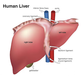
Human Liver

Although liver cells called hepatocytes are sometimes used for liver transplants, they can't be generated in the lab as easily as stem cells can. Researchers transplanted stem cells into mice with severely damaged livers, finding that major areas of the liver had regrown over several months and were improved the organ's performance.

This is the first time that researchers have been able to cause an organ in a living animal to regenerate using stem cells. If human stem cells behave in a way similar to the mice cells, they said, transplanting stem cells -- or using drugs to motivate a patient's own liver to produce stem cells and regenerate itself -- could replace liver transplants.

"This research has the potential to revolutionize patient care by finding ways of co-opting the body's own resources to repair or replace damaged or diseased tissue," said Dr. Rob Buckle, director of science programs for the U.K.'s Medical Research Council, in a press release.

Source : http://goo.gl/Fi95NQ

Sunday 19 July 2015

Scientists Created Mini Heart on Microchip With Stem Cells

Scientists belonging to the University of California of the USA conducted a research that led to the development of mini heart (Cardiac Microphysiological System) on a microchip using human stem cells. The study was led by Indian-origin scientist Anurag Mathur.

The Cardiac Microphysiological System, which is hardly the width of a human hair, is expected to replace non-human animal models that are used in drug discovery and development process.


The invention was published in the second week of March 2015 in the Scientific Reports journal in an article titled Human iPSC-based Cardiac Microphysiological System For Drug Screening Applications.

What is Microphysiological System?

Microphysiological systems are engineered organs that are developed to address the formidable pharmacological and physiological gaps between monolayer cell cultures, animal models and humans. The Cardiac Microphysiological System is the latest human organ - after a lung, a liver and a piece of intestine – were developed under laboratory environment.

How Cardiac Microphysiological System/ mini heart was developed?

It was developed using human-induced pluripotent stem cells (iPSC) that can form many different types of tissues. These cells, once tricked into forming heart tissue, were grown around a special silicon microchip with cell and media channels that mimicked the heart’s blood vessels.

Significance of Cardiac Microphysiological System

Apart from replacing the animal models used in the drug discovery process that do not mimic human responses; the organ-on-chip will help in the development of personalized medicine in future as doctors will be able to predict how certain drugs react on specific patients, thus preventing many illnesses and loss of valuable time.

Doctors will be able to calculate the approximate dose needed for patients with heart conditions by deploying this bionic heart technology as they will be able to have his or her heart modelled in a lab with all the tests done.

Thursday 16 July 2015

Researchers Create Model Of Early Human Heart Development From Stem Cells

Researchers at the University of California, Berkeley, in collaboration with scientists at the Gladstone Institutes, have developed a template for growing beating cardiac tissue from stem cells, creating a system that could serve as a model for early heart development and a drug-screening tool to make pregnancies safer.

In experiments to be published Tuesday, July 14, in the journal Nature Communications, the researchers used biochemical and biophysical cues to prompt stem cells to differentiate and self-organize into micron-scale cardiac tissue, including microchambers.

"We believe it is the first example illustrating the process of a developing human heart chamber in vitro," said Kevin Healy, a UC Berkeley professor of bioengineering, who is co-senior author of the study with Dr. Bruce Conklin, a senior investigator at the Gladstone Institute of Cardiovascular Disease and a professor of medical genetics and cellular and molecular pharmacology at UC San Francisco. "This technology could help us quickly screen for drugs likely to generate cardiac birth defects, and guide decisions about which drugs are dangerous during pregnancy."

To test the potential of the system as a drug-screening tool, the researchers exposed the differentiating cells to thalidomide, a drug known to cause severe birth defects. They found that at normal therapeutic doses, the drug led to abnormal development of microchambers, including decreased size, problems with muscle contraction and lower beat rates compared with heart tissue that had not been exposed to thalidomide.

"We chose drug cardiac developmental toxicity screening to demonstrate a clinically relevant application of the cardiac microchambers," said Conklin. "Each year, as many as 280,000 pregnant women are exposed to drugs with evidence of potential fetal risk. The most commonly reported birth defects involve the heart, and the potential for generating cardiac defects is of utmost concern in determining drug safety during pregnancy."

The new milestone comes nearly four months after Healy and other UC Berkeley researchers publicly debuted a system of beating human heart cells on a chip that could be used to screen for drug toxicity. However, that heart-on-a-chip device used pre-differentiated cardiac cells to mimic adult-like tissue structure.

In this new study, the scientists mimicked human tissue formation by starting with stem cells genetically reprogrammed from adult skin tissue to form small chambers with beating human heart cells. Conklin's lab at Gladstone, an independent, nonprofit life science research organization affiliated with UC San Francisco, supplied these human induced pluripotent stem cells for this study.

The undifferentiated stem cells were then placed onto a circular-patterned surface that served to physically regulate cell differentiation and growth.

By the end of two weeks, the cells that began on a two-dimensional surface environment started taking on a 3D structure as a pulsating microchamber. Moreover, the cells had self-organized based upon whether they were positioned along the perimeter or in the middle of the colony.

Compared with cells in the center, cells along the edge experienced greater mechanical stress and tension, and appeared more like fibroblasts, which form the collagen of connective tissue. The center cells, in contrast, developed into cardiac muscle cells. Such spatial organization was observed as soon as the differentiation started. Center cells lost the expression of octamer-binding transcription factor 4 (OCT4) and epithelial cadherin (E-cadherin) faster than perimeter cells, which are critical to the development of heart tissue.

"This spatial differentiation happens in biology naturally, but we demonstrated this process in vitro," said study lead author Zhen Ma, a UC Berkeley postdoctoral researcher in bioengineering. "The confined geometric pattern provided biochemical and biophysical cues that directed cardiac differentiation and the formation of a beating microchamber."

Could eventually replace animal models

Modeling early heart development is difficult to achieve in a petri dish and tissue culture plates, the study authors said. This area of study has typically involved the dissection of animals at different stages of development to study the formation of organs, and how that process can go wrong.

"The fact that we used patient-derived human pluripotent stem cells in our work represents a sea change in the field," said Healy. "Previous studies of cardiac microtissues primarily used harvested rat cardiomyocytes, which is an imperfect model for human disease."

The researchers pointed out that while this study focused on heart tissue, there is great potential for use of this technology to study other organ development.

"Our focus here has been on early heart development, but the basic principles of patterning of human pluripotent stem cells, and subsequently differentiating them, can be readily expanded into a broad range of tissues for understanding embryogenesis and tissue morphogenesis," said Healy.

source : http://goo.gl/BfdN7j

Tuesday 14 July 2015

Stem Cells Provide Lasting Pain Relief in Mice

Chronic pain caused by the nerve damage of type 2 diabetes, surgical amputation, chemotherapy and other conditions is especially intractable because it resists painkilling medications. 

But in a study on mice, a Duke University team has shown that injections of stem cells from bone marrow might be able to relieve this type of neuropathic pain. The researchers say their findings, which appear July 13 in the Journal of Clinical Investigation, may also advance cell-based therapies in chronic pain conditions, lower back pain and spinal cord injuries. 

The team used a type of stem cell known as bone marrow stromal cells (BMSCs), which are known to produce an array of healing factors and can be coaxed into forming most other types of cells in the body. 

Stromal cells are already being tested in small-scale clinical studies of people with inflammatory bowel disease, heart damage and stroke. They have also shown promise for treating pain. However, it’s not clear how they work. 

“Based on these new results, we have the know-how and we can further engineer and improve the cells to maximize their beneficial effects,” said Ru-Rong Ji, professor of anesthesiology and neurobiology in the Duke School of Medicine.

In his team’s study, the researchers used stromal cells to treat mice with pain caused by nerve damage. They delivered the cells by a lumbar puncture, infusing them into the fluid that bathes the spinal cord. 

Mice treated with the bone marrow stromal cells were much less sensitive to painful stimuli after their nerve injury compared with the untreated mice, the researchers found. 

“This analgesic effect was amazing,” Ji said. “Normally, if you give an analgesic, you see pain relief for a few hours, at most a few days. But with bone marrow stem cells, after a single injection we saw pain relief over four to five weeks.”

Pictures of the animals’ spinal cords showed that the injected stem cells had set up shop alongside the nerve cells in the spinal cord.

To understand how the stem cells alleviated pain, the researchers measured levels of anti-inflammatory molecules that had been previously linked to pain, finding that one in particular, TGF-β1, was present in higher amounts in the spinal fluid of the stem cell-treated animals compared with the untreated animals. 

TGF-β1 is a protein that is typically secreted by immune cells and is common throughout the body. Research has shown that people with chronic pain have too little TGF-β1, Ji said.

In the new study, chemically neutralizing TGF-β1 reversed the pain-killing benefit of the BMSCs, suggesting that the secretion of this protein was a major reason why the cells helped with pain. Injecting TGF-β1 directly into spinal cord fluid provides relief too, but only for a few hours, Ji said.

By contrast, bone marrow stromal cells stay on site for as much as three months after the infusion, the scientists found. This is the right length of time, Ji said, because if the stem cells stayed permanently there could be risk of them becoming cancerous.  

Importantly, BMSCs also migrate to the site of injury. That’s because a molecule emitted from the injured nerve cells called CXCL12 -- which has also previously been linked to neuropathic pain -- acts as a homing signal of sorts, attracting the stem cells. 

The next step will be to find a way to make the stromal cells more efficient. “If we know TGF-β1 is important, we can find a way to produce more of it,” Ji said. In addition, the cells may produce other pain-relieving molecules; Ji’s group is working to identify those. 

source : http://goo.gl/IIBCdD

Thursday 9 July 2015

Researchers Develop Patient-Specific Heart Cells From Stem Cells

Induced pluripotent stem cells (IPSC) could be the key to the future of personalized medicine, as a new study published by Stanford Cardiovascular Medicine has successfully used the stem cells to recreate patient-specific heart cells. Their research could carve the path for other areas of medicine to use IPSC, which can be transformed into any body cell, to develop patient-specific treatments for any disease.
Published on June 18, the study advances the general quest in the medical community to develop personalized medicine using IPSC, which are easy to find and can be taken from the skin of patients. The transformed cells in the study seemed as if they came directly from the patient’s heart itself, explained lead author and postdoctoral scholar HaoDi Wu. Wu said the heart cells were even beating in the petri dish.

Stem Cell Treatment for Heart Disease

IPSC are preferred by scientists as they carry specific DNA of patients and can be used to treat patient-specific diseases, such as dilated cardiomyopathy (DCM), a common heart condition caused by genetic mutations. The researchers’ study focused specifically on looking at DCM mutations within a single family.
Although they did discover the specific mutation that caused DCM in one family of patients as well as a compound to treat the disease, the significance of their experiment lay in the method.  Their novel use of IPSC to model human cells allowed them to narrow the target for finding the DCM mutations in the whole human genome.
Senior author of the paper Joseph Wu, director of the Stanford Cardiovascular Institute and professor of medicine and of radiology, described how their results highlight the potential of IPSC. Today, doctors often go through trial and error to find the right drugs for their patients; IPSC would allow them to test drugs on stem cells first, not the patients themselves.
“We’re hoping that in the future, instead of you being the guinea pig, it’ll be patient-specific, IPS-cell-derived brain cells, derived heart cells, derived kidney cells, liver cells in a dish,” Joseph Wu said. “In essence, you’re doing a clinical trial in a dish.”
The study was funded by the American Heart Association (AHA) and the National Institutes of Health. Joseph Wu received the Established Investigator Award from the AHA for funding, and according to AHA project coordinator Micah Moughon, the best applications for funding are those that accomplish the mission stated on their website: “to build healthier lives free of cardiovascular diseases and stroke.”
“People have been working for a very long time, fighting for cardiomyopathy,” HaoDi Wu said. “This will be the initial step – we take advantage of the IPSC cardiomyocyte system to find the detailed mechanism of the single-gene mutation in cardiomyopathy.”
While the study narrows down the target for finding mutations that cause cardiomyopathy, it cannot guarantee that the mutation exists in all patients. The researchers explained that their team has a long way to go before they can even consider the possibility of a new drug or bringing IPSC to current medicine.
“Before you get that goal of doing clinical trial in a dish or doing personalized medicine 10, 20 years from now, you [have] got to do these experiments to show the feasibility [of IPSC],” Joseph Wu said. “There are other patients with other mutations that cause cardiomyopathy. So the question is, ‘Does this finding also hold true for other mutations?’ And does this finding also hold true for patients without mutations but also heart failure?”
The project already had years of research behind it when it started – research on the possibility of even creating IPSC, on using them in experiments. Although the lab spent about two years of work to find the mutation for only one family, as the experiment is reviewed and tested more, it will be easier to narrow down and streamline the process to apply to all kinds of patients and not just in cardiology.
“This is a novel and unprecedented approach to diagnosing disease, physiology,” said Tzung Hsiai, friend of Joseph Wu and cardiology professor at UCLA. “The ramification is not limited to cardiomyopathy disease but has relation to many other diseases. We will be able to predict pharmacological treatments – for laymen’s terms, drug treatments and patient’s responses to medication.”
Joseph Wu explained that there is a widespread interest to develop personalized medicine and incorporate it into the industry. There is still a long way to go but most areas of medicine are taking strides toward this concept and are hopeful for the future, he said.
“We just happened to focus in cardiology because I’m a cardiologist; it’s a cardiac lab, and we’ve been doing this for a while,” Joseph Wu said. “Other labs are focused on using the same set up to address Parkinson’s disease,  Alzheimer’s disease.”
“[It’s a] general movement toward using these IPS cells for disease modeling, understanding the disease, drug discovery and for personalized medicine,” he added.


Source : http://goo.gl/FRESfa

Monday 6 July 2015

BBC PRESENTER WITH LEUKAEMIA FINDS LAST-MINUTE STEM CELL DONOR

The award-winning journalist diagnosed with leukaemia has found a donor after a the BBC launched an appeal on her behalf.

Sue Lloyd Roberts tweeted on Sunday that she had found a match and would return to hospital in two weeks for a stem cell transplant to cure her aggressive form of the disease.

The BBC hosted an open day at New Broadcasting House on 22 June to encourage potential donors to come forward.


Her initial donor was found to be unsuitable at the last moment forcing her back to square one.

Roberts, 62,  was first diagnosed when she collapsed at the Majorca hotel she runs with her husband, BBC producer Nick Guthrie in January.

After tests at University College London hospital she underwent chemotherapy while she waited for a stem cell donor.

She is currently in remission but needs the donor to cure her of the disease completely. Tests have shown her white cell platelets are too low for her to attempt another round of chemotherapy and it was now "a race against time".

She told the Daily Mail: “I have been mired in hell for six months.

“The kind of leukaemia I have means I need a stem cell transplant to survive. It is vital. I was mightily annoyed when I discovered what I had because I have always been a fitness fanatic. But I believe in getting on with it.”

Writing in the Independent, Lloyd-Roberts said the disease had forced her to put her life on hold as she could not work. She described her experience with chemotherapy: "I found the incarceration hard to bear. I have always been an active person. "

Source : http://goo.gl/q3PKZ7